

The other half of your heart
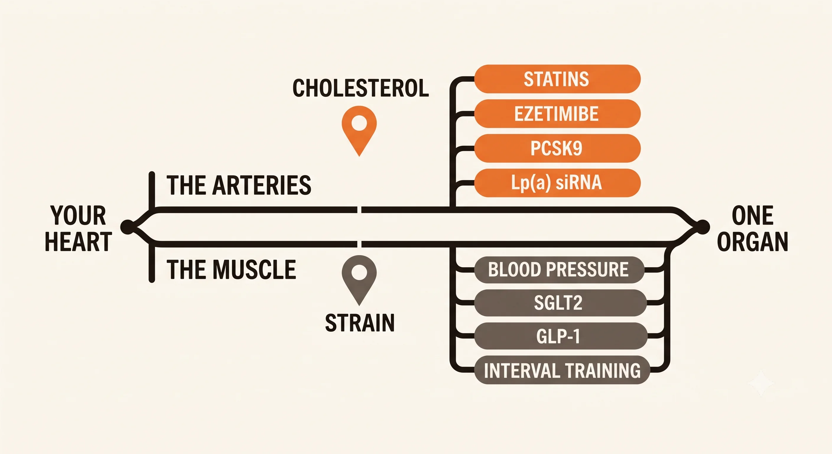
If you've spent the last few years reading about prevention, you can probably tell me your ApoB. You might know your Lp(a), and you'll have a view on whether it matters. You've read about statins and had opinions about the arguments.
But from my conversations with patients, it seems like another axis has been covered more superficially or potentially not at all, and if you've been reading the same sources it has probably passed you by too. What you've learned is about the plumbing, the arteries that feed the heart. None of it is about the pump. And the pump fails on its own schedule, for its own reasons, in people whose arteries are in fine shape.
That's not something you've ignored. It's that the prominent advocates of lipid health know about lipids. A cardiologist spends their career on the whole organ, and the muscle half rarely makes it into the same discourse.
Your heart has two ways of failing and you've only been taught one
| The arteries: what you already know | The muscle: this piece | |
|---|---|---|
| The measurement that matters | ApoB | Global longitudinal strain |
| What it improves on | LDL cholesterol | Ejection fraction |
| The imaging | Calcium score, CT angiogram | Strain echocardiography, cardiac MRI |
| The blood test with a catch | Lp(a), measured once | NT-proBNP, low if you're heavy |
| What drives the damage | Decades of particle exposure | Decades of pressure, plus inflammation |
| First-line treatment | A statin | Blood pressure control |
| The lifestyle lever | Diet | Structured exercise |
| The newer drugs | PCSK9 inhibitors, Lp(a) siRNA | SGLT2 inhibitors, GLP-1 RA, finerenone |
| The supplement question | Fish oil, plant sterols | CoQ10, resveratrol |
| How you follow it | Repeat the lipid panel | Repeat the echo, retest VO2max |
The plumbing problem is the one you know. Plaque builds in the coronary arteries, blood flow drops, and at some point you get angina or a heart attack. ApoB drives it, and it's measurable, treatable and trackable.
The pump problem is different. The muscle gets stiffer. A stiff heart fills poorly, and filling is half the job. The heart has to relax and take blood in before it can push any out. When it can't fill properly, pressure backs up into the lungs. You get short of breath climbing stairs. Your blood pressure swings more than it used to. And your ejection fraction, the figure everyone checks, stays completely normal the whole time.
The muscle stiffens from load and from age, and the two compound
Two things drive it, and they compound.
The first is load. Every time the heart contracts against a higher pressure, it has to work harder, and muscle that works harder gets thicker. That's not a disease process, it's the same adaptation you'd get in any muscle. But a thicker ventricle is a less compliant one, and years of blood pressure that's been a little too high is the commonest way to get there. A wall that grew to meet a load doesn't relax the way it used to.
The second is aging itself. Mitochondrial dysfunction is one of the recognised hallmarks of aging, and mitochondria turn out to do more than make energy. When they start failing, they leak reactive oxygen species and fragments of their own DNA into the cell, and those fragments trip the same alarm systems that respond to infection. The result is a low-grade inflammatory state, and in the heart that inflammation drives fibrosis. Collagen accumulates between the muscle cells. The tissue gets stiffer for reasons that have nothing to do with your arteries.
Put those together and you have a heart that's thicker from decades of pressure and stiffer from decades of low-grade inflammation, in someone whose lipid panel is immaculate.
There's a name for where you probably are, and it isn't heart failure
Cardiologists stage heart failure from A to D, and in 2022 the American Heart Association and American College of Cardiology renamed the early ones. Stage B is now officially called pre-HF, and the criteria are short enough to read, because you may recognise yourself in them.
Stage B means no symptoms and no signs of heart failure, plus any one of the following:
- Structural heart disease: ventricular hypertrophy, chamber enlargement, reduced systolic function,
- Evidence of raised filling pressures, either measured with catheters or suggested by Doppler on an
- Risk factors plus a raised natriuretic peptide, or a persistently raised cardiac troponin, with no
Look at that list against the person described at the start. A thickened ventricle from years of blood pressure is ventricular hypertrophy. An enlarged left atrium is chamber enlargement. A high E/e' is noninvasive evidence of raised filling pressures. And reduced strain is on the list by name, which is the answer to anyone who tells you strain is a research measurement.
Any one of those, on its own, with no symptoms at all, is pre-HF. Not heart failure. Not a diagnosis you need to be alarmed about. But a recognised stage with a name, and the guideline's stated aim at this stage is to treat the risk and the structural change to prevent the syndrome from developing.
That's the stage we're going to focus on and it's where a surprising number of well-informed healthy-feeling people already are. If you're over 60, carrying some extra weight, and you've had blood pressure that's been treated a bit but never quite tightly, you're the profile.
Stage C is when symptoms show up. The whole point of knowing about Stage B is that it's upstream of that, and it's where the useful decisions live.
A stiff heart reads normal at rest and gives out on exertion
Here's something that explains a lot of missed diagnoses. A stiff heart can look completely normal at rest. Lying on an examination couch, your heart has all the time in the world to fill, and the pressures inside it are fine.
Exercise removes that buffer. Your heart rate goes up, which means less time to fill on each beat, and the blood coming back from your legs increases. A compliant ventricle absorbs that easily. A stiff one can't, so the pressure required to fill it climbs steeply, and that pressure backs into the lungs. That's the shortness of breath, and it's why it shows up on the second flight of stairs and not while you're sitting down.
As a field, cardiology took a long time to recognize that passive measurements can't pick up this dysfunction. But we can now measure this directly, putting catheters in the heart while people exercise and comparing the readings against the echocardiogram at the same moment. It's the reason a resting scan that reads normal doesn't settle the question in someone who gets breathless walking uphill.
A stiff system turns small changes into large pressure swings
This one almost never gets explained, and it's the sign patients most often bring up.
The same process that stiffens the ventricle tends to stiffen the arteries, and the two together change how your blood pressure behaves. A compliant system absorbs a change in volume or vessel tone with only a small change in pressure. A stiff system doesn't. The same extra half-litre of fluid, the same surge of adrenaline, now moves the pressure much further. So readings that used to sit in a narrow band start bouncing, 118 one morning, 165 the next afternoon, and you get told to check your cuff.
The cuff is usually fine. When this was measured directly with pressure-volume catheters, people with a stiff heart and preserved ejection fraction had both a stiffer ventricle and stiffer arteries than people the same age with the same blood pressure. The lability isn't noise in the measurement. It's a mechanical consequence of the stiffening this piece is about.
That study was small, and it explains a mechanism. But swinging readings have other causes and most of them are commoner than this one. But if your pressures have become erratic and nobody's been able to tell you why, this belongs on the list.
Ejection fraction measures the squeeze, not the filling
Ejection fraction is the percentage of blood the heart pushes out with each beat. It's on every echocardiogram, and a normal one closes the conversation.
But EF only measures the squeeze. A stiff heart that can't fill still squeezes out a normal fraction of whatever it managed to take in. So EF stays normal while the muscle is quietly changing, and it stays normal right up until things are quite far along. We don't need to blame EF for failing to do this job. But it's just answering a different question from the one you're asking.
Strain falls before anything else does
There's a better measurement, but many routine echocardiograms don't include it unless it's explicitly asked for. It's called global longitudinal strain, or GLS. Instead of measuring how much blood came out, it measures how much the muscle itself shortens. It is the actual deformation of the tissue.
Strain can turn abnormal while EF is still normal. That's the entire reason to care about it.
A GLS below 16% suggests the muscle isn't working normally. Only about 2.8% of healthy people fall below that. Normal sits around 21%.
Two influences lower it that aren't disease. Age nudges it. People over 60 average about 20.0% against 21.0% under 60. And your own blood pressure lowers it, independently of any damage. So an untreated hypertensive reads a little lower for reasons that are about load, not injury.
One practical note. If you're going to follow strain over time, get it done in the same place on the same equipment. And a change smaller than about 2.5 strain units isn't necessarily a change, it's inside the measurement error. A GLS that goes from 19.5 to 18.0 might be due to chance but if it continues to drop, it likely is a real change.
You can score most of this yourself before anyone scans you
There's a validated score for the probability of this kind of heart failure, called the H2FPEF score. It was built by measuring pressures directly in the heart, which makes it unusually well grounded. Six items:
| Points | |
|---|---|
| BMI over 30 | 2 |
| On two or more blood pressure medications | 1 |
| Atrial fibrillation, paroxysmal or persistent | 3 |
| Age over 60 | 1 |
| Estimated pulmonary artery pressure over 35 mmHg (echo) | 1 |
| E/e' over 9 (echo) | 1 |
Seven of those nine points don't need an echocardiogram. You know your BMI, your age, how many blood pressure pills you take and whether you've ever been in atrial fibrillation. So you can get most of the way to an answer at your kitchen table, and the scan settles the last two points.
Now the important caveat, and it's the kind of thing that gets left out. This score was built in people who already had unexplained breathlessness and were sent for invasive testing, a group where 64% turned out to have the condition. In that setting the odds roughly double for every point. But the probabilities printed alongside the score don't transfer to someone who feels well. The scale bottoms out at 20%, and an asymptomatic 45-year-old scoring zero doesn't have a one-in-five chance of heart failure.
So use it the way it's useful. A high score in someone who feels fine isn't a diagnosis. It's a reason to actually look. And if you're 62 and your BMI is 31, you're at 3 points before anything else is counted, which tells you the score is picking up how common this profile is, not that you're ill.
The measurements behind those criteria, and the job each one does
Left atrial volume index. The left atrium sits upstream of the stiff ventricle and takes the pressure first. It enlarges slowly, over years, which makes it a good long-run record of what the heart has been living with. It also means a flat value is a good result, not a disappointing one.
E/e'. An estimate of filling pressure from the echo. It's one of the two points on the score above.
Left ventricular mass and wall thickness. The direct measure of how much muscle years of pressure have built. It's on the same scan.
NT-proBNP. A blood test for the strain the heart wall is under, and in the patient this piece is about, it's the laboratory value most likely to reassure you when it shouldn't. Carrying extra weight pushes it down, so an obese person can have a normal-looking result and still have the problem.
In a pooled analysis of nearly 15,000 people, those with a BMI of 35 or above hit a given level of actual risk at an NT-proBNP of 158 pg/mL, while people below that BMI didn't reach the same risk until 450, roughly three times higher for identical danger. At one commonly used cutoff, the real event rate ran from 3.5 per 100 person-years in people under a BMI of 30 up to 7.3 in those at 40 or above. The same result means about twice the risk at the heavy end.
So the test is useful, and it's least useful in exactly the person most likely to need it. If you're heavy and short of breath and your NT-proBNP came back fine, that result hasn't ruled much out.
Peak VO2. Your maximum oxygen uptake, and the single best measure of what your cardiovascular system can actually deliver. Of everything on this list it's the most repeatable, it needs no radiation, and it's the one you can most clearly improve.
DXA body composition. Cheap, low-dose and repeatable, and it separates fat from lean in a way the scale can't. Visceral fat is the compartment that tracks with this problem, and it's the first to fall when things go well.
The stiffening responds to treatment
Everything above is diagnosis. This part matters more, because unlike a lot of what gets discussed in prevention, the stiffening is not a one-way process.
The trial that showed the stiffening reverses
Sedentary, healthy, middle-aged people were randomised to two years of supervised exercise training, or to a control group doing stretching and balance work. Not a few weeks. Two years. And the researchers measured ventricular stiffness properly, with catheters in the heart, building pressure-volume curves before and after.
The stiffness constant fell from 0.072 to 0.051 in the training group and didn't move in the controls (0.0635 to 0.062). Peak VO2 rose 18%, from 29.0 to 34.4, while the control group drifted slightly down. The trained hearts took in more blood for the same filling pressure, which is the mechanical definition of a more compliant ventricle.
Fifty-three people completed it, so it's a small study. But it measured ventricular stiffness directly instead of inferring it, and it answers the question that matters: this is modifiable in exactly the group this piece is about, at exactly the age they are.
One caveat the authors were careful about. This was middle age. The same group's earlier work suggests the window doesn't stay open indefinitely, and starting at 70 after decades of sitting is a different proposition from starting at 52.
Blood pressure control has the strongest evidence behind it
If you take one thing from this, take this one. In a trial of over 9,000 people, treating systolic blood pressure to a target below 120 instead of below 140 cut new heart failure by nearly 40%, hazard ratio 0.62, with a confidence interval from 0.45 to 0.84. Death from cardiovascular causes fell by a similar margin, 0.57 (0.38 to 0.85).
Heart failure was one of the outcomes most strongly reduced by tighter blood pressure control. That's the single best-evidenced thing on this page, and it's aimed squarely at the mechanism described earlier, less load, less hypertrophy, less stiffening.
There's a trade-off and you should know it. Intensive treatment brought more low blood pressure, more electrolyte problems, more kidney injury and more fainting. That's a conversation to have, not a reason to skip it, and it's why the target is a discussion and not a rule.
Two practical points. Base decisions on an averaged series of home readings, never on the one alarming reading that sent you to the doctor, a single high reading tends to fall on its own at the next check, which makes anything you did in between look effective. And if your readings swing, that's a reason to measure more often, not less.
Exercise, and specifically what kind
The trial above used a structured programme built around aerobic training, including interval work, with sessions increasing over the first months and then sustained. The dose mattered and so did the duration. This isn't a walking-more intervention.
Add resistance training. The reason is in the next section, and it's one of the few places where the newer weight-loss drugs and the exercise story interact.
Weight, and the drugs everyone's asking about
GLP-1 receptor agonists have been tested in people who already have this condition with obesity, and they improved symptoms and the ability to exercise. Those trials enrolled patients further along than you, so they don't directly answer whether starting one at Stage B prevents Stage C. Nobody has run that trial.
What they do tell you is that the obesity-driven version of this disease responds to taking the weight off, which is a reasonable thing to know when you're deciding what to do about your own.
Then there's the muscle question, which comes up in every conversation about these drugs. Yes, lean mass falls. The evidence says that's an adaptive response, not damage. People carrying more weight carry more muscle to move it, and weight loss by any route, surgery, diet, drugs, takes some of it back. What matters is whether function follows, and it doesn't seem to. In one study using MRI, psoas muscle volume fell 9.3% over 24 weeks while the fat inside the muscle didn't change, chair-rise time and walking speed both edged in the better direction, and the proportion of people with a slow walking speed dropped from 63% to 46%.
There's a cardiac analogy that makes this intuitive. Left ventricular hypertrophy develops in proportion to the pressure the heart works against, and treating the blood pressure makes it regress. Nobody calls that regression damage, the muscle grew to meet a load and shrank when the load lifted. Skeletal muscle in someone losing 20 kilos is doing the same thing.
Where it does need attention is in older and frailer people, where there's less reserve to give up. That's a question of who's a good candidate, not a reason to avoid the drug, and it's the argument for pairing it with resistance training instead of skipping it.
If you have type 2 diabetes, two more options open up
This is the part most people don't know. SGLT2 inhibitors and finerenone are available on their diabetes and kidney indications, independently of any heart problem. And both have been shown to help in preserved ejection fraction heart failure.
So if you're the patient this piece describes and you also have type 2 diabetes, you're not choosing between treating the diabetes and protecting the muscle. The choice of which agent to use for the diabetes can take the heart into account, and that's a conversation to have with whoever manages it.
To be clear about what's claimed: no trial has tested these drugs for preventing heart failure in someone at Stage B. The argument is that when you already qualify for a drug class on other grounds, its effects on the heart are a legitimate input into which one you pick.
The mitochondrial supplements, and how far the evidence actually goes
Go looking for heart-muscle supplements and you'll land on the mitochondrial ones. CoQ10, resveratrol, urolithin A, the NAD precursors. The pitch is built on the mechanism from earlier in this piece, and that mechanism is sound. Failing mitochondria do drive inflammation, that inflammation does contribute to fibrosis, and it's a recognised hallmark of aging. This isn't fringe biology.
The trials are more mixed than either the marketing or the debunking suggests, so here's what each one actually did.
Resveratrol, in exactly this patient
This is the closest anyone has come to testing a supplement in the person this piece describes. Eighty hypertensive patients were assigned to 400 mg a day of resveratrol on top of their usual treatment, or to usual treatment alone, for six months. Here is what each measurement did, because the individual estimates matter far more than whether the study called them significant.
| After six months, adjusted | Usual care | Resveratrol | Difference |
|---|---|---|---|
| E/e′ (filling pressure) | 12.10 ± 0.36 | 11.09 ± 0.33 | −1.0 |
| Strain, GLS (%) | −19.80 ± 0.25 | −20.51 ± 0.23 | −0.71 |
| Left atrial diameter (cm) | 3.72 ± 0.05 | 3.60 ± 0.04 | −0.12 |
| PICP, a fibrosis marker | 1043.8 ± 32.5 | 878.1 ± 35.1 | −166 |
| Galectin-3, a fibrosis marker | 10.76 ± 0.18 | 10.13 ± 0.20 | −0.63 |
Every one of those moves in the direction you'd want. Left ventricular structure didn't differ between the groups, and arterial stiffness fell within the resveratrol group but not compared with control.
Now put the strain result next to a number from earlier in this piece. The measurement error on GLS is about 2.5 strain units. The resveratrol group's strain differed from control by 0.71. That's less than a third of what a single scan can resolve. Averaged across forty people the difference is detectable, but in one person, on one machine, it would be invisible. The same applies to a left atrium 1.2 millimetres smaller.
That isn't a reason to dismiss the study. It's a reason to be precise about what it supports: a consistent nudge in the right direction across several independent measurements, each too small to see in an individual over six months. What would make it convincing is the same nudge sustained over years, which is the timescale you actually care about and the one no supplement trial has run.
The design limits matter too. There was no placebo, the comparison group carried on with usual care and everyone knew who was taking what, which counts for more than usual when the endpoints are echo measurements that need human interpretation. Eighty people, one centre, six months.
So it's the most on-target result in this space, at a dose you can buy, and it argues for a proper blinded trial before a purchase.
Selenium with CoQ10, and the longest follow-up of anything here
Four hundred and forty-three elderly Swedes took selenium and CoQ10 together, or placebo, for four years, then were followed for twelve years with nobody lost. Cardiovascular mortality stayed lower in the supplemented group, hazard ratio 0.58, 95% confidence interval 0.42 to 0.79. Cardiovascular death reached 38.7% in the placebo group.
The twelve-year follow-up is why this one earns your attention, since the horizon is the whole problem with supplement evidence. But read what it does and doesn't tell you. It measured deaths, not the muscle. It says nothing about whether strain, filling pressure or atrial size moved, so it can't tell you what to expect on your own panel.
The population is the real caveat. The paper states plainly that selenium intake is low across Europe, and this was a rural Swedish community. Correcting a deficiency is a different proposition from supplementing someone already replete, and selenium intake in the United States is generally adequate. You also can't tell which of the two components did anything, because they were only ever given together.
Urolithin A, where the primary endpoint gated everything behind it
Sixty-six adults aged 65 to 90, mean age 72, took 1,000 mg of urolithin A or placebo. The trial pre-specified an ordered hypothesis: ATP production would only be tested confirmatorily if the six-minute walk distance separated first. It didn't, so everything downstream is exploratory by the trial's own design, which is a more useful thing to know than any single result.
Here are the muscle endurance figures. Contractions until fatigue rose by 95.3 (SD 115.5) on urolithin A against 11.6 (SD 147.4) on placebo in the hand muscle, and 41.4 (65.5) against 5.7 (127.1) in the shin. Those are large central estimates sitting inside spreads wider than the effect, which is what a small study of a variable measurement looks like. Plasma acylcarnitines, ceramides and C-reactive protein all fell relative to placebo.
It's a signal, in skeletal muscle, not the heart, from a study that didn't clear its own first gate.
The mechanism is sound and the evidence is thin, both at once
Read together, these say something more useful than either the marketing or a debunking. The measurements mostly move the right way, and every effect is small enough that six months can't distinguish it from nothing. That's not a scandal. It's what you'd expect from interventions acting on a process that takes decades, tested over a couple of quarters.
Which is exactly why the question to ask about any of them isn't whether a trial reached significance. It's whether the effect is plausible, whether anything suggests harm, and whether you can measure your own trajectory well enough to tell in five years. Something biologically plausible, with no harm signal, bought with your own money, is a reasonable choice made with your eyes open. What it isn't is established.
There's also one place where the mitochondrial story has a well-tested answer, and it isn't a supplement. The exercise trial earlier in this piece is the best-evidenced mitochondrial intervention available to you, and one of the approved diabetes drugs raises the heart's energy production directly. The biology these products are sold on is sound. What reliably improves it is in the sections above.
The one supplement tested for this specifically, and it failed
The supplement most often marketed for heart-muscle health is dietary nitrate, usually as beetroot concentrate, on the reasoning that it boosts nitric oxide and should improve exercise capacity.
It's been tested properly and it doesn't. In a double-blind crossover trial across 17 sites, inhaled inorganic nitrite changed peak oxygen consumption by −0.20 mL/kg/min, with a confidence interval from −0.56 to 0.16. That's not a study that was too small to find an effect. The interval is tight around zero, which is a different and more informative result than a wide one. Daily activity, quality of life, filling pressure and NT-proBNP were all similarly flat. A separate trial of an oral nitrate found no benefit either, and activity levels were slightly worse.
The limit on that conclusion: those trials ran weeks to months in people who already had the condition. They close the question of whether nitrate helps established disease. They don't formally close the question of what dietary nitrate does over thirty years, because that trial will never be run. But they do remove the specific promise the supplements are sold on.
Where this leaves you
If you've optimised your lipids, you've done work that counts, and none of it is wasted. You've also been working on one half of the organ.
The other half is measurable with tools that already exist, most of it from one echocardiogram ordered by someone who knows to ask for strain, a blood test read against your body size, and a walk on a treadmill with a mask on. And what it measures responds. Blood pressure control has the strongest evidence behind it, two years of structured training reversed the stiffening in people your age, and the weight side has more options than it did five years ago.
None of that is a reason for alarm. Stage B is not a diagnosis of heart failure, and most people reading this who score points on that table will never develop it. It's a reason to look, once, at a part of your heart nobody's shown you.
References
What pre-heart-failure is
- Heidenreich PA, et al. 2022 AHA/ACC/HFSA guideline for the management of heart failure. Circulation. 2022;145:e895–e1032. https://doi.org/10.1161/CIR.0000000000001063 — Stage B / pre-HF definition, Table 3.
Why the muscle stiffens
- López-Otín C, Blasco MA, Partridge L, Serrano M, Kroemer G. Hallmarks of aging: an expanding universe. Cell. 2023. https://doi.org/10.1016/j.cell.2022.11.001
- Tracy E, Rowe G, LeBlanc AJ. Cardiac tissue remodeling in healthy aging: the road to pathology. Am J Physiol Cell Physiol. 2020;319:C166–C182. https://doi.org/10.1152/ajpcell.00021.2020
Measuring it
- Obokata M, Reddy YNV, Borlaug BA. Role of diastolic stress testing in the evaluation for heart failure with preserved ejection fraction. Circulation. 2017;135:825–838. https://doi.org/10.1161/CIRCULATIONAHA.116.024822
- Kawaguchi M, Hay I, Fetics B, Kass DA. Combined ventricular systolic and arterial stiffening in patients with heart failure and preserved ejection fraction. Circulation. 2003;107:714–720. https://doi.org/10.1161/01.CIR.0000048123.22359.A0. Correction: Circulation. 2020;141:e809. https://doi.org/10.1161/CIR.0000000000000778
- D'Elia N, et al. Normal global longitudinal strain: an individual patient meta-analysis. JACC Cardiovasc Imaging. 2020;13(1). https://doi.org/10.1016/j.jcmg.2019.07.020
- Balinisteanu A, et al. Vendor differences in 2D-speckle tracking global longitudinal strain: an update on a 10-year standardization effort. Eur Heart J Cardiovasc Imaging. 2025;26:1360–1373. https://doi.org/10.1093/ehjci/jeaf155. See also the ASE/EACVI clinical consensus statement on strain echocardiography, 2025. https://doi.org/10.1016/j.echo.2025.07.007
- Reddy YNV, Carter RE, Obokata M, Redfield MM, Borlaug BA. A simple, evidence-based approach to help guide diagnosis of heart failure with preserved ejection fraction. Circulation. 2018;138:861–870. https://doi.org/10.1161/CIRCULATIONAHA.118.034646
- Ostrominski JW, et al. Natriuretic peptides, body mass index, and clinical outcomes in heart failure with mildly reduced or preserved ejection fraction. JACC. 2025;86:1823–1839. https://doi.org/10.1016/j.jacc.2025.08.028
What changes it
- Howden EJ, et al. Reversing the cardiac effects of sedentary aging in middle age — a randomized controlled trial. Circulation. 2018;137:1549–1560. https://doi.org/10.1161/CIRCULATIONAHA.117.030617
- SPRINT Research Group. A randomized trial of intensive versus standard blood-pressure control. N Engl J Med. 2015;373(22). https://doi.org/10.1056/NEJMoa1511939. Final report: https://doi.org/10.1056/NEJMoa1901281
- Anker SD, et al. Empagliflozin in heart failure with a preserved ejection fraction (EMPEROR-Preserved). N Engl J Med. 2021. https://doi.org/10.1056/NEJMoa2107038. Solomon SD, et al. Dapagliflozin in heart failure with mildly reduced or preserved ejection fraction (DELIVER). N Engl J Med. 2022. https://doi.org/10.1056/NEJMoa2206286. Solomon SD, et al. Finerenone in heart failure with mildly reduced or preserved ejection fraction (FINEARTS-HF). N Engl J Med. 2024;391(16). https://doi.org/10.1056/NEJMoa2407107
- Kosiborod MN, et al. Semaglutide in patients with heart failure with preserved ejection fraction and obesity (STEP-HFpEF). N Engl J Med. 2023;389(12). https://doi.org/10.1056/NEJMoa2306963. Packer M, et al. Tirzepatide for heart failure with preserved ejection fraction and obesity (SUMMIT). N Engl J Med. 2025;392(5). https://doi.org/10.1056/NEJMoa2410027
- Linge J, Birkenfeld AL, Neeland IJ. Muscle mass and glucagon-like peptide-1 receptor agonists: adaptive or maladaptive response to weight loss? Circulation. 2024;150:1288–1298. https://doi.org/10.1161/CIRCULATIONAHA.124.067676
- Ditzenberger GL, et al. Effects of semaglutide on muscle structure and function in the SLIM LIVER study. Clin Infect Dis. 2025;80(2):389–396. https://doi.org/10.1093/cid/ciae384
- Verma S, et al. Empagliflozin increases cardiac energy production in diabetes. JACC Basic Transl Sci. 2018. https://doi.org/10.1016/j.jacbts.2018.07.006
Supplements
- Zheng X, et al. Effects of resveratrol supplementation on cardiac remodeling in hypertensive patients: a randomized controlled clinical trial. Hypertens Res. 2023;46:1493–1503. https://doi.org/10.1038/s41440-023-01231-z
- Alehagen U, Aaseth J, Alexander J, Johansson P. Still reduced cardiovascular mortality 12 years after supplementation with selenium and coenzyme Q10 for four years. PLoS ONE. 2018;13(4):e0193120. https://doi.org/10.1371/journal.pone.0193120
- Liu S, et al. Effect of urolithin A supplementation on muscle endurance and mitochondrial health in older adults: a randomized clinical trial. JAMA Netw Open. 2022. https://doi.org/10.1001/jamanetworkopen.2021.44279
- Borlaug BA, et al. Effect of inorganic nitrite vs placebo on exercise capacity among patients with heart failure with preserved ejection fraction: the INDIE-HFpEF randomized clinical trial. JAMA. 2018. https://doi.org/10.1001/jama.2018.14852
Want your heart muscle actually measured?
Book a Video Visit